MULTIMODALITYADAPTIVE IMAGING SYSTEM FOR EQUINE EXTREMITY STUDIES
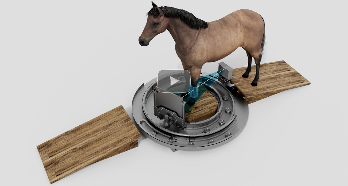
STAY IN FOCUS ON THE ANATOMY DETAILS OF YOUR REAL INTEREST
Adapting FOV to a desired extremity location without moving the animal(by source and detector motion modulation)Registering and compensating of animal motion in order to obtain high spatial resolution images. No need for deep anesthesia, just light sedatives are required.Extremity radiography and fluoroscopy. CT X-Ray techniques:40-120 kV / 0.1-0.3 mAs per frameCT scanning time:15 sec (expedited mode) or 30 sec(high quality mode)CT FOV:D270xH270 mmIsotropic 3D spatial resolution 300 micronPatient database with full DICOM support and PACS compatibility.